Tansen complex, sirmour chouraha, below LIC Office, Rewa, Madhya Pradesh 486001 +91 810976 5307 +91 87709 57521 07662 – 465792

Digital X- Ray

Digital X- Ray

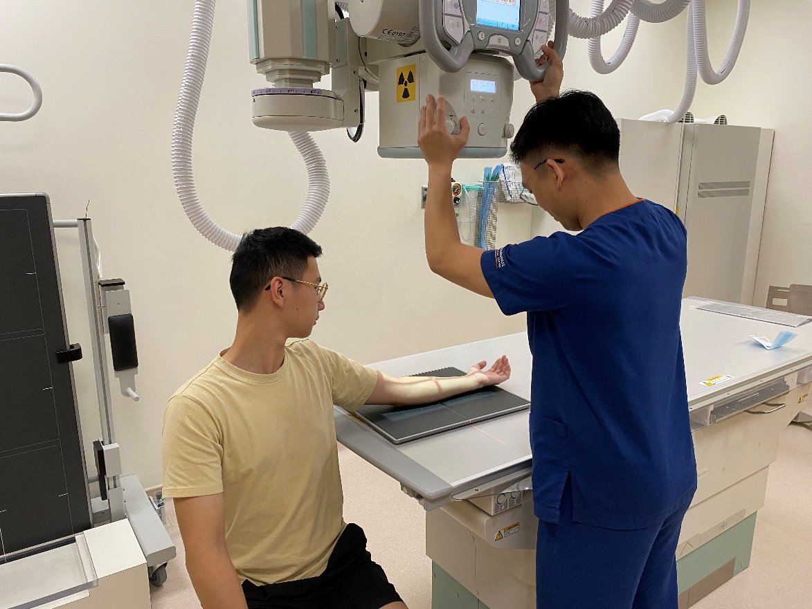
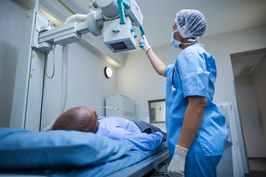
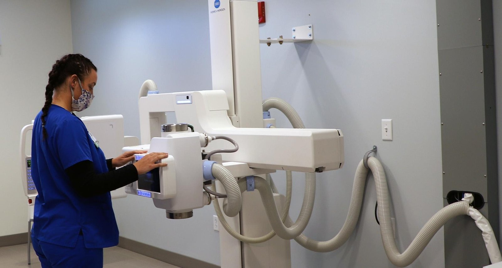
A Digital X-Ray is a form of X-ray imaging where digital X-ray sensors are used instead of traditional photographic film. This technology has significantly transformed diagnostic imaging, offering advantages in terms of speed, efficiency, and the reduction of radiation exposure for patients. Digital X-rays are used across various medical fields, including dentistry, mammography, and orthopedics, among others.

Components of Digital X-Ray Systems

  • X-Ray Tube: Generates the X-rays.
  • Digital Detector: Captures the X-rays after they pass through the patient’s body. There are two main types of digital detectors:
  • Direct Digital Radiography (DR) Detectors: Convert X-ray photons directly into electrical signals.
    Computed Radiography (CR) Detectors: Use a phosphor plate to capture X-ray images, which are then read into a computer that converts the image into a digital format.
  • Computer System: Processes and stores the digital images. Advanced software allows for image enhancement, zooming, and other manipulations to aid in diagnosis.

Advantages of Digital X-Rays

  • Reduced Radiation Exposure: Digital X-ray systems require less radiation to produce an image of similar contrast to traditional radiography.
  • Instant Image Availability: The images are available for viewing immediately after exposure, eliminating the wait time associated with film development.
  • Image Enhancement: Digital images can be enhanced and manipulated for better clarity and detail, aiding in more accurate diagnoses.
  • Easy Storage and Sharing: Digital images can be stored in digital medical records, making it easier to retrieve and share with other healthcare professionals for consultation or comparison over time.

Applications

Digital X-rays are used in various diagnostic applications, including:

  • Dentistry: To detect cavities, inspect the tooth roots, check the health of the bone around the teeth, and monitor the general health of the teeth and jawbone.
  • Chest Radiography: To examine the lungs, heart, and chest wall.
  • Orthopedics: To view fractures, dislocations, and the detection of orthopedic implants.
  • Mammography: For the early detection of breast cancer.

Safety and Considerations

While digital X-ray systems reduce radiation exposure, any level of radiation poses a risk. Therefore, it’s essential to use these systems judanitiously and only when medically necessary. Protective measures, such as lead aprons, may be used to shield parts of the body not being imaged from exposure.

Principles of Operation

Digital X-ray systems utilize a digital image capture device to record and store the X-ray images electronically. When X-rays pass through the body, they are absorbed at varying levels by different tissues, depending on the density and composition of those tissues. The digital sensors capture this pattern to create a detailed image that represents the internal structure of the area being examined.

In conclusion, digital X-ray technology represents a significant advancement in medical imaging, offering improvements in safety, efficiency, and diagnostic capability. Its ability to provide instant, high-quality images with lower doses of radiation has made it an invaluable tool in modern medicine.

Get directions

Find us on map

Contact & Appointment

royaldentalclinicbhopal@gmail.com

Emergency

+91 99778 4111